视频第一部分原理内容(红色为不确定部分):
Initial studies in the photo elastic techniques seem to have been in 1937 with Highdman and hush. But photo elastic visualization of ultrasound was first practically developed by Doctors Hence and White in UK around 1968. In the 1970’s it was popularized by Dr King hall at British area.
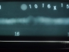
The image shown is provided by Dr hence shows a multiple exposure of 4 MHz pulse refracted at 45 degrees. The principle is relatively simple, pulse light is passing through the light source at a fix repetition frequency on the order of 100 Hz. Unpolarised light from the spark gap passes through a polarizing filter. This changes the light that pass through the polarizer to linear polarized light.
This is followed by a quarter wave plate that changes the linear polarized light to circular polarized light, a second quarter wave plate and a analyzer are inserted into the light path, and the orientation can figured to none of the light passing through the analyzer.
A clear transparent solid such as glass is inserted in the light path, between the quarter wave plates. When stress in the glass are set up by mechanical disturbing such as ultrasonic pulse. It causes a change the rotation of polarized light, change the stress along the light path change the conditions that none of the light passing through the analyzer.
So a small amount of light can then pass through the observer on the other side of the analyzer. by adding a delay circuit, we can adjust the position along the sound path that is illuminated by the pulse, and the camera allows the observer to record the image.
希望能帮上忙,另外,关于光弹部分内容可以给您介绍清华的一位老师,他对这研究颇深。
杨彬 2013 12 31
电话:01089438696
mail:yangbin@higgsndt.com
|
 [复制链接]


 提升卡
提升卡 置顶卡
置顶卡 沉默卡
沉默卡 喧嚣卡
喧嚣卡 变色卡
变色卡 千斤顶
千斤顶 显身卡
显身卡